
|
Biomedical Engineering Course Assoc. prof. Yoshihisa YAMAOKA E-mail:yamaoka [at] cc.saga-u.ac.jp (Please replace [at] with @ in e-mail address.) Academic Staff Database |
 |
|
|||||||||||||||||||||||||||||||||
Research Field : Biomedical imaging, Photoacoustic imaging, Femtosecond laser applications, Ultrafast nonlinear optics, Optical interferometry
Membership in Academic Societies :
Secretary of Professional Group of Photoacoustic Imaging, Councilor of Japan Society of Histochemistry and Cytochemistry (JSHC), Technical committee member of Japan Society for Laser Microscopy, Member of Optical Society of America (OSA), Member of Society of Photo-Optical Instrumentation Engineers (SPIE), Member of Japanese Society of Applied Physics (JSAP), Member of Japanese Society for Medical and Biological Engineering (JSMBE), Member of the Japan Society of Ultrasonics in Medicine
Secretary of Professional Group of Photoacoustic Imaging, Councilor of Japan Society of Histochemistry and Cytochemistry (JSHC), Technical committee member of Japan Society for Laser Microscopy, Member of Optical Society of America (OSA), Member of Society of Photo-Optical Instrumentation Engineers (SPIE), Member of Japanese Society of Applied Physics (JSAP), Member of Japanese Society for Medical and Biological Engineering (JSMBE), Member of the Japan Society of Ultrasonics in Medicine
 Photoacoustic imaging to visualize deep structures in living tissues
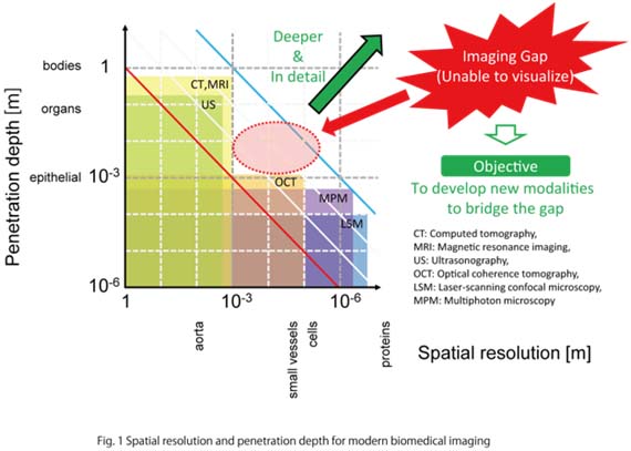
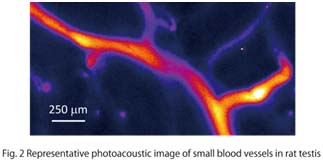
Accurate detection of the growth extent of cancer cells in depth direction is very important. In particular, depth discrimination of melanoma cells is extremely essential because even a tiny difference in the invasion of cancer cells strongly determines the outcome for the patients. Depth resolution less than 10 μm (size of cells) is required to discriminate individual cells. As for the imaging depth, several mm is required, because the thickness of skin tissues is of several-mm order. Imaging with 10-μm resolution is also important to visualize small vascular channels, key constituents in living tissues. In this way, imaging over several-mm depth with 10-μm spatial resolution is desired in clinical applications.Optical imaging typified by laser-scanning microscopy has a spatial resolution high enough to observe single cells. However the penetration depth of more than 1 mm is difficult to obtain. On the other hand, magnetic resonance imaging (MRI), computed tomography (CT) and ultrasonography (US), which are commonly used in medicine, can visualize deeply enough to investigate whole human bodies. However, the spatial resolution is not high enough to observe single cells. Thus, imaging modalities with a spatial resolution of 10 μm at around several mm depth do not exist (Fig. 1). To bridge this gap in imaging modalities, we have studied and developed various types of photoacoustic imaging (especially, two-photon photoacoustic microscopy (TP-PAM, Fig. 2).
 |
 |
 |
 |