
|
Biomedical Engineering Course Prof. Eiji TAKAHASHI E-mail:eiji [at] cc.saga-u.ac.jp (Please replace [at] with @ in e-mail address.) Academic Staff Database Laboratory Home Page |
 |
|
||||||||||||||||||||||||||||||||||||||||||
Research Field : biomedical engineering
Membership in Academic Societies :
International Society on Oxygen Transport to Tissue(Executive Committee Member), International Society on Oxygen Transport to Tissue(ISOTT2015 Scientific Comittee), International Society on Oxygen Transport to Tissue(41st Annual ISOTT Meeting: Scientific Advisory Committee), International Society on Oxygen Transport to Tissue, International Society on Oxygen Transport to Tissue, International Society on Oxygen Transport to Tissue, International Society on Oxygen Transport to Tissue, American Physiological Society, Mitochondrial Physiology Society
International Society on Oxygen Transport to Tissue(Executive Committee Member), International Society on Oxygen Transport to Tissue(ISOTT2015 Scientific Comittee), International Society on Oxygen Transport to Tissue(41st Annual ISOTT Meeting: Scientific Advisory Committee), International Society on Oxygen Transport to Tissue, International Society on Oxygen Transport to Tissue, International Society on Oxygen Transport to Tissue, International Society on Oxygen Transport to Tissue, American Physiological Society, Mitochondrial Physiology Society
His specialty is Physiology and Biomedical Engineering. He is also a Physiology Educator certified by The Physiological Society of Japan since 2015.
BIOIMAGINGS are powerful tool to explore the function of biomolecules in individual cells. We are particularly interested in the OXYGEN because this molecule is not only crucial for energy production in mitochondria but also mediates a range of genetic responses for survival. OXYGEN is particularly important in cancer because the molecule defines the biological characteristics of tumor cells. Using sophisticated techniques to visualize oxygen dynamics and other metabolic processes in cells, we are working to elucidate the mechanism for survival of cancer cells in hypoxic environment.
 Bioimaging of cellular oxygen transport and metabolism
We are developing novel techniques for imaging oxygen transport to and within cells. Using these techniques, we have demonstrated mechanisms by which cells undergo necrotic cell death even at physiological oxygen concentrations. Details can be found at www.ee.saga-u.ac.jp/sensor/Res_ET/O2_imaging.html.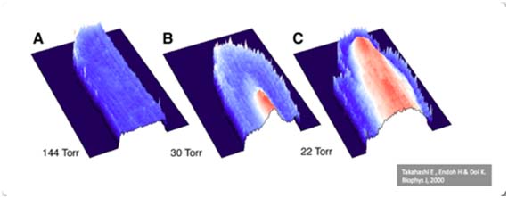
A life with less oxygen?
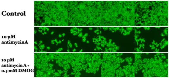
Recently, we have demonstrated that some cancer cell lines can sustain mitochondrial membrane potential without oxygen when cellular oxygen sensor (PHD/HIF-1) is activated. Find more inTakahashi E and Sato M: Anaerobic respiration sustains mitochondrial membrane potential in prolyl hydroxylase pathway-activated cancer cell line in a hypoxic microenvironment. Am J Physiol Cell Physiol. 2014 Feb 15;306(4):C334-42.
Cancer cells prefer higher oxygen?
To elucidate the initial mechanisms of hematogenous metastasis of cancer cells, we hypothesized that cancer cell might migrate towards regions with higher oxygen concentration such as intratumor micro vessels along the gradient of oxygen concentration. To test this hypothesis, we have devised the gap cover glass (GCG) by which oxygen concentration gradients were established in monolayer cultured cells. We demonstrated a directional migration of MDA-MB-231 cells under the oxygen gradient.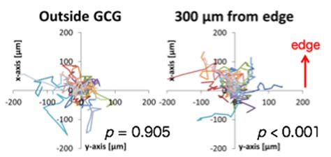
 |
 |
 |
 |